Using dual-energy x‐ray imaging to enhance automated lung tumor tracking during real-time adaptive radiotherapy
Abstract
Purpose: Real-time, markerless localization of lung tumors with kV imaging is often inhibited by ribs obscuring the tumor and poor soft-tissue contrast. This study investigates the use of dual-energy imaging, which can generate radiographs with reduced bone visibility, to enhance automated lung tumor tracking for real-time adaptive radiotherapy.
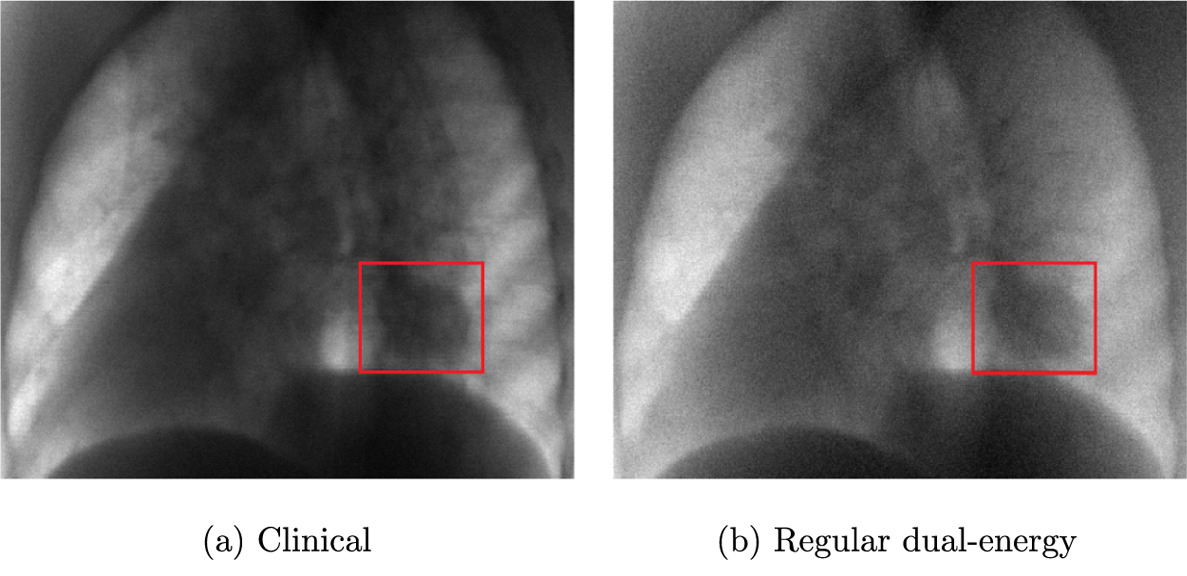
Methods: LkV images of an anthropomorphic breathing chest phantom were experimentally acquired and radiographs of actual lung cancer patients were Monte-Carlo-simulated at three imaging settings: low-energy (70 kVp, 1.5 mAs), high-energy (140 kVp, 2.5 mAs, 1 mm additional tin filtration), and clinical (120 kVp, 0.25 mAs). Regular dual-energy images were calculated by weighted logarithmic subtraction of high- and low-energy images and filter-free dual-energy images were generated from clinical and low-energy radiographs. The weighting factor to calculate the dual-energy images was determined by means of a novel objective score. The usefulness of dual-energy imaging for real-time tracking with an automated template matching algorithm was investigated.
Results: Regular dual-energy imaging was able to increase tracking accuracy in left–right images of the anthropomorphic phantom as well as in 7 out of 24 investigated patient cases. Tracking accuracy remained comparable in three cases and decreased in five cases. Filter-free dual-energy imaging was only able to increase accuracy in 2 out of 24 cases. In four cases no change in accuracy was observed and tracking accuracy worsened in nine cases. In 9 out of 24 cases, it was not possible to define a tracking template due to poor soft-tissue contrast regardless of input images. The mean localization errors using clinical, regular dual-energy, and filter-free dual-energy radiographs were 3.85, 3.32, and 5.24 mm, respectively. Tracking success was dependent on tumor position, tumor size, imaging beam angle, and patient size.
Conclusions: This study has highlighted the influence of patient anatomy on the success rate of real-time markerless tumor tracking using dual-energy imaging. Additionally, the importance of the spectral separation of the imaging beams used to generate the dual-energy images has been shown.